
 23 Tumanyan St, Yerevan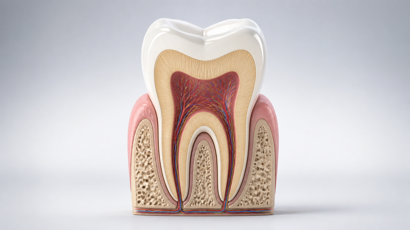
Key points in one minute
The main thing: a tooth is a living structure, and not just one solid material
- Enamel covers the crown and protects it, but is not restored by the body like skin; acid exposure and plaque increase the risk of tooth decay.
- Dentin makes up the bulk of the tooth and is associated with sensitivity: when it is exposed or a lesion approaches it, reactions to cold, hot and sweet things may become more noticeable.
- The pulp contains nerves and blood vessels; its inflammation or damage may require endodontic evaluation rather than self-treatment with pain medications.
- The root, periodontal ligament, cementum and bone support the tooth; bleeding gums, mobility or swelling are reasons not to postpone the visit.
Tooth map in simple words: crown, neck and root
The part visible above the gum is called the crown. The narrow transition at the edge of the gum is conventionally called the neck, and the root is located in the alveolar bone of the jaw. For different groups of teeth, the shape and number of roots differ: the incisors mainly bite off, the canines hold and tear food, the premolars and molars grind it. The group name describes the function and position, but does not replace the evaluation of a specific tooth.
On the chewing teeth, the upper surface has tubercles and grooves; in the front teeth, the cutting edge and the surface on the lip side are important. The doctor may talk about the occlusal, buccal, lingual or palatal surfaces, contact between teeth and occlusion. Occlusion is the way the upper and lower teeth fit together. It affects load distribution, but does not in itself explain any pain or clicking in the jaw.
- The crown is the part of the tooth above the gum.
- The root is the part in the bone that helps hold the tooth in place.
- The canal is a narrow space within the root where the pulp tissue passes.
- Periodontium is the tissue that connects the root to the surrounding bone.
Enamel, dentin and pulp: what each layer does
Enamel is the outer mineralized layer of the crown. It is very hard, but can lose minerals under the influence of acids that form in plaque with the frequent intake of free sugars. At the beginning, the process may look like a dull white spot and not hurt. Regular brushing with a fluoride toothpaste and limiting frequent sugar-sweetened snacks help reduce the risk, but home cleaning will not close an existing cavity.
Beneath the enamel lies dentin, a less hard tissue penetrated by microscopic tubules. It forms the main volume of the crown and root. If dentin is exposed due to decay, wear, cracked or receding gums, cold, hot, sweet or touch may cause a brief reaction. A similar complaint occurs for different reasons, so it is impossible to decide based on one symptom that a specific type of treatment is needed.
In the center is the pulp complex: soft tissue with nerves, vessels and cells. At the root, it continues into the root canal and communicates with the surrounding tissues through an opening at the apex. Deep caries, a crack, trauma or severe inflammation can affect the pulp. Prolonged spontaneous pain, pain when biting or discoloration of a tooth is a reason to consult a dentist; the need for root canal treatment is determined by clinical tests and images, and not just by the patient’s perception.
| Part | Where is | What is important to the patient |
|---|---|---|
| Enamel | Outside of the crown | Protects the surface; the initial changes may not hurt. |
| Dentin | Under enamel and cement | Associated with sensitivity when exposed or affected. |
| Pulp | Inside the crown and canals | Contains blood vessels and nerve fibers; a doctor is assessing her condition. |
| Cement and periodontium | On and around the root | They help attach the tooth to the bone and respond to inflammation or stress. |
Root, cementum, periodontium and periodontium: what holds the tooth
The root is covered with cementum, a hard tissue to which the fibers of the periodontal ligament are attached. This ligament connects the tooth to the alveolar bone, absorbs normal loads and transmits the sensation of pressure. Therefore, a healthy tooth does not “fuse” with the bone directly. On an x-ray, the doctor evaluates not only the root, but also the surrounding bone, the periodontal ligament space and the apical area.
The word periodontium is broader: it refers to the tissues surrounding and supporting the tooth, including the gums, periodontal ligament, cementum and bone. Plaque and inflammation of the gums may initially appear as bleeding, redness, or an unpleasant odor. As the disease progresses, it can affect supporting tissues. Do not attribute repeated bleeding to a hard brush and do not try to treat mobility with folk remedies - you need an examination and professional assessment.
How to read terms in the treatment plan and in the image
The number of a tooth is a way to accurately indicate its position in the dentition; numbering systems vary, so it is helpful to ask your doctor to show a specific tooth on a diagram or picture. The terms "caries", "pulpitis", "periodontitis", "periapical tissue", "resorption" or "fissure" describe different findings and should not be interpreted independently. One image also does not answer all questions: it is compared with complaints, examination, gum condition and pulp vitality tests.
A close-up photo clearly shows an individual tooth and the area around its root; a panoramic examination gives a general overview of the jaws. Three-dimensional visualization is not prescribed “just in case,” but when the expected benefit for resolving the issue exceeds the need for additional research. Ask what problem the shot is intended to solve, what alternatives are available, and how the result will change the plan. This is especially important if treatment is offered only on the basis of photographs, correspondence, or unclear descriptions of symptoms.
- Which tooth and which surface is causing the complaint?
- What exactly is visible upon examination and in the picture, and what is still an assumption?
- Do I need observation, restoration, root canal treatment, or consultation with another specialist?
- What signs mean you shouldn't wait until your next scheduled visit?
What might be normal and when do you need a doctor?
Brief sensitivity after extreme cold or heat does not always indicate a serious problem: it may be due to exposed dentin, a recent procedure, or another condition. But a recurring symptom deserves evaluation, especially if it gets worse or interferes with eating. It is not normal for self-monitoring to include persistent night or throbbing pain, a visible cavity, chipping, increasing swelling, purulent discharge, tooth trauma, noticeable discoloration or mobility.
If there is rapidly growing swelling of the face or neck, fever with severe deterioration in well-being, difficulty breathing or swallowing, emergency medical attention is required according to local regulations. Do not apply aggressive substances to the gums and do not start antibiotics or other medications based on advice from the Internet. The article helps formulate questions, but does not replace diagnosis or determine the urgency of an individual case.
Save before your visit
Questions worth asking your dentist
- What part of the tooth or surrounding tissue is most likely related to my complaint?
- What examination and imaging findings confirm the diagnosis, and which require observation?
- Is it possible to save tooth tissue and what treatment or restoration options exist?
- What symptoms after the visit require an unscheduled visit?
Sources
- Oral health — World Health Organization
- What Is a Root Canal? — American Association of Endodontists
- Tooth decay — American Dental Association


