
 23 Tumanyan St, Yerevan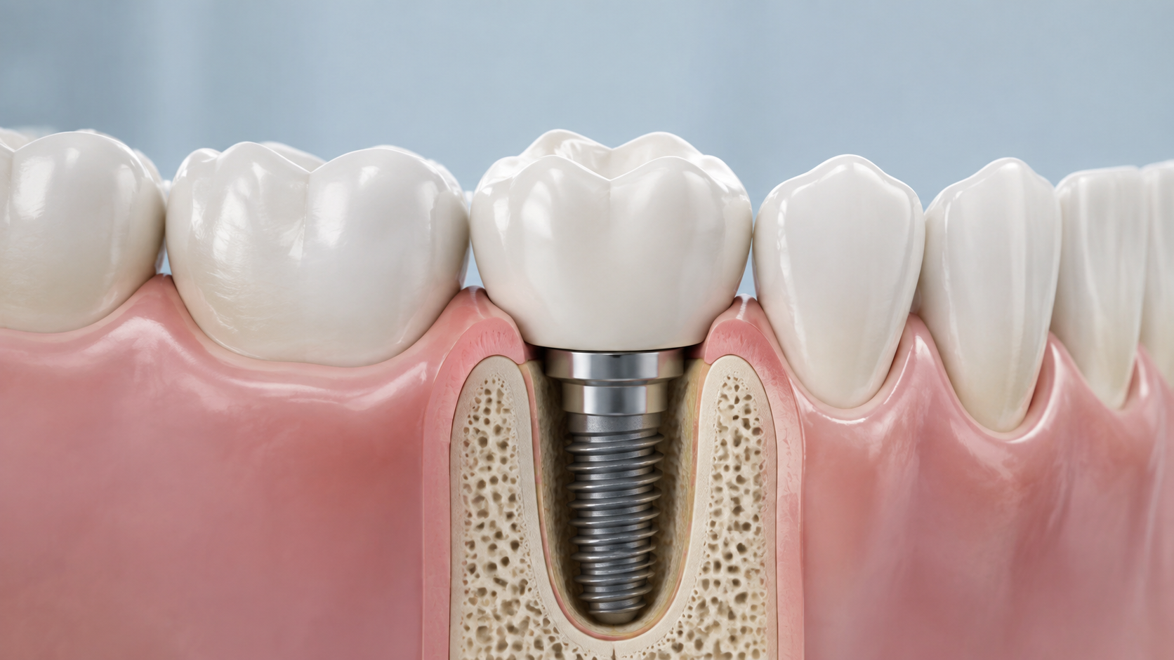
Key points in one minute
Key point: inflammation around an implant cannot be assessed reliably at home; an early examination helps choose the safest next step
- An implant has no periodontal ligament, so its surrounding tissues are assessed differently from those around a natural tooth.
- Repeated bleeding with gentle cleaning, swelling, tenderness, discharge or a change in contour are reasons to see a dentist.
- Assessment may include history, plaque and restoration review, gentle probing and, when indicated, X-rays; no single sign makes a diagnosis.
- Treatment depends on the cause, severity and condition of bone and soft tissue; home care matters but does not replace professional treatment.
What soft tissue around an implant means
Soft tissue includes the gum and mucosa surrounding an implant-supported crown. Its thickness, width and contour can affect cleaning access, comfort and appearance. There is no single ‘normal width’ for everyone: implant position, bone volume, smile line, neighbouring teeth, crown shape and individual anatomy all matter.
Tissues change while healing after implant placement, a healing abutment or a crown. Timelines and sensations differ, so another person's experience is not a safe guide. A dentist considers more than colour: plaque, bleeding on examination, access for cleaning, tissue levels, the stability of components and, when needed, bone changes on an image are reviewed together.
- The contour around a crown should allow gentle, effective cleaning.
- Keratinised gum may be considered during planning, but its presence alone does not determine implant success or failure.
- A visible change in the gumline is not always inflammation, but its cause deserves assessment.
Symptoms and risk factors that matter
Signs worth discussing with a dentist include recurrent bleeding when cleaning, redness or swelling, pain, a feeling of pressure, discharge, bad breath or taste, exposed parts of the restoration and a changed crown position. Peri-implant mucositis is inflammation of the soft tissue without confirmed supporting-bone loss; peri-implantitis involves inflammation together with bone loss. These conditions cannot be distinguished without examination and baseline information.
Risk may be higher with biofilm accumulation, smoking, past or active periodontitis, a restoration that is difficult to clean around, retained cement and irregular maintenance visits. These factors do not mean that a problem will certainly develop and do not explain every symptom. Do not remove a crown yourself, use harsh products or start antibiotics without assessment: this can obscure important signs and complicate care.
| What you notice | Why it matters | A practical next step |
|---|---|---|
| Bleeding during cleaning or examination | It can indicate soft-tissue inflammation | Arrange an examination while continuing gentle cleaning |
| Pain, swelling, discharge or an unpleasant taste | Inflammation and other complications should be ruled out | Contact a dentist soon |
| Rapidly increasing swelling, fever, difficulty swallowing or breathing | This may be urgent | Seek urgent in-person medical care |
How a dentist identifies the cause
Assessment begins with your history: when the implant and crown were placed, what has changed, gum disease, smoking, health conditions, earlier treatment and previous imaging. The dentist then examines tissue and restoration, reviews cleaning access, plaque, bleeding, possible component mobility and may gently measure relevant findings around the implant. The result is interpreted with baseline records and the rest of the mouth.
An X-ray may be useful to assess the bone level or compare it with prior images. It does not show soft tissue fully and does not replace a clinical examination. The cause may instead be crown shape, retention, bite forces, recession or another issue. That is why remote advice and one symptom are not a sound basis for choosing treatment.
Treatment, soft-tissue grafting and limits
When soft-tissue inflammation is confirmed, a dentist first addresses conditions that retain biofilm: home cleaning may be refined, accessible surfaces professionally cleaned, and the restoration checked for shape or retained material that prevents hygiene. If bone loss is suspected, care can become more complex and may include non-surgical and surgical stages. The method, materials and outlook depend on the defect and individual factors.
Soft-tissue grafting or augmentation may sometimes be discussed to improve tissue thickness, contour, cleaning comfort or appearance. It is not automatically needed for every implant and does not rule out later tissue changes. Whether it is considered before implant placement, at placement or after the crown depends on anatomy and the treatment goal. Monitoring, reshaping a restoration or another treatment plan may be alternatives; each option needs a discussion of risks, limits and maintenance.
- Ask which specific finding is being addressed and how the result will be assessed.
- Ask whether comparative imaging is needed and what alternatives exist.
- Agree on a maintenance schedule after active treatment is complete.
Home care and when not to wait
At home, continue gentle cleaning as instructed by your dentist, with a soft toothbrush and suitable interdental aids. The size of an interdental brush, floss, water flosser and frequency depend on the restoration: an oversized aid can traumatise tissue, while an undersized one may clean poorly. Do not try to mask bleeding with frequent strong solutions or stop hygiene altogether.
Book an appointment for recurring bleeding, swelling, pain, discharge, unpleasant taste, a change in gum level or difficulty cleaning. Rapid facial swelling, fever, severe pain, difficulty swallowing or breathing need urgent in-person medical care. If you are unsure in Yerevan, arrange a consultation: the dentist can combine symptoms, examination and imaging if it is genuinely needed, without promising an outcome.
- Do not try to unscrew, remove or refit a crown yourself.
- Bring previous images and the implant-placement date if available.
- Ask to be shown a safe cleaning technique for your specific restoration.
Save before your visit
Questions worth asking your dentist
- Do my signs suggest mucositis, peri-implantitis or another cause?
- Is imaging needed to compare the current bone level with baseline records?
- Does the shape of my crown or bridge make cleaning difficult?
- Which home-care routine and review interval suit my restoration?
Sources
- Peri-implant diseases and conditions: consensus report — European Federation of Periodontology
- Academy of Osseointegration and American Academy of Periodontology consensus — American Academy of Periodontology
- Outcomes of peri-implantitis treatment followed by supportive care — International Team for Implantology


