
 23 Tumanyan St, Yerevan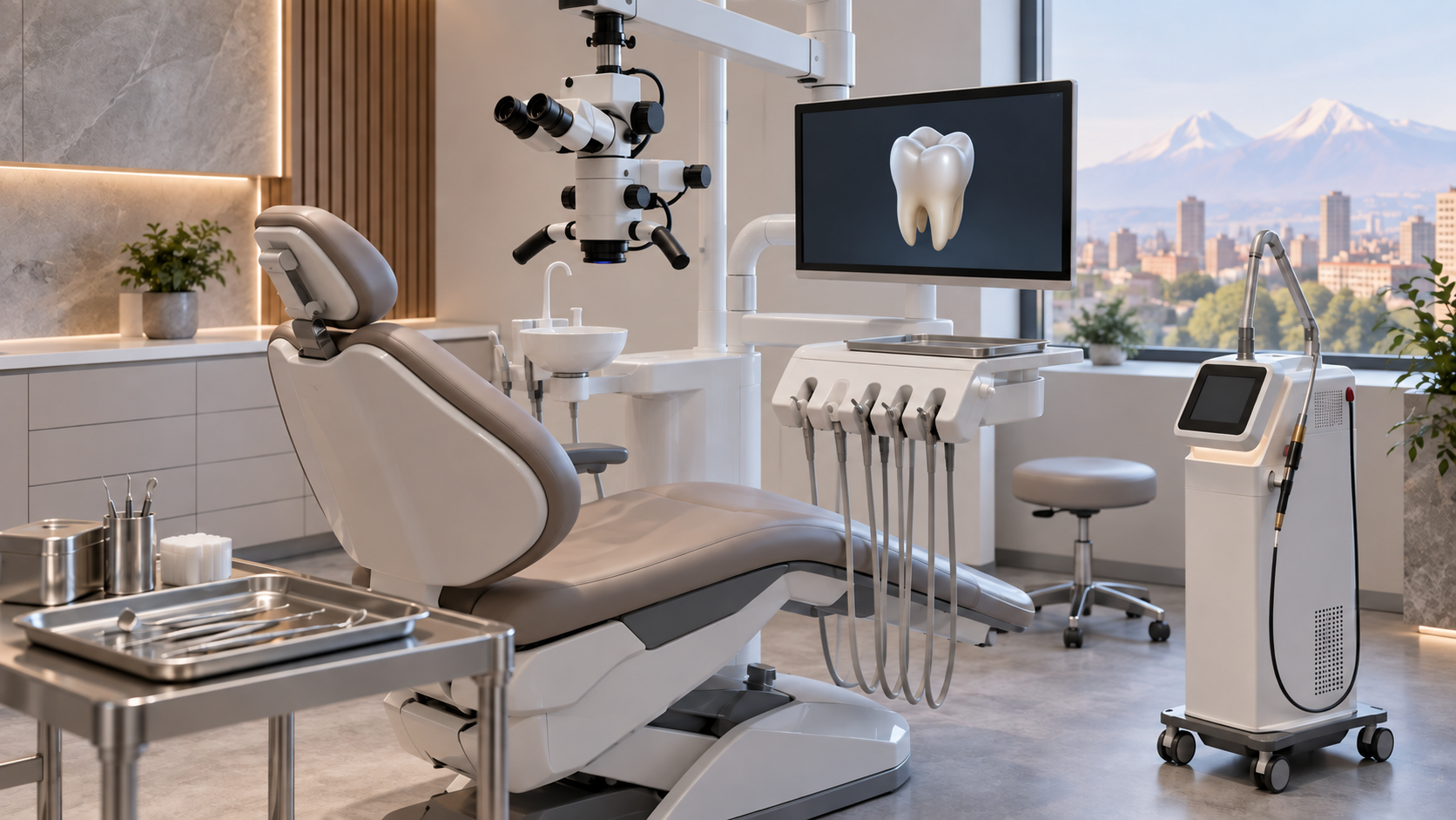
Key points in one minute
Before you choose a technology
- Start with a diagnosis and a clear treatment goal; select the tool to suit the task.
- A microscope, surface scanner and 3D X-ray examination provide different information and are not interchangeable.
- Laser benefits depend on the procedure, appropriate settings and protection; they do not apply to every treatment.
- A digital preview can help discuss a plan, but an image of a future smile is not a promised result.
From a concern to the right tool
People seek care for pain, a damaged or missing tooth, or a change to their smile. The dentist first reviews health and treatment history, examines the mouth and decides what information is needed. This determines whether imaging, magnification, scanning or another tool is appropriate. Technologies may complement one another, but every patient does not need a complete digital package.
Separate data collection, clinical decision-making and the procedure itself. A scanner records tooth shape, the dentist assesses whether restoration is feasible, and digital design supports manufacturing. An error in the first stage can carry forward. An impressive screen image does not establish that a tooth is healthy or that a proposed restoration is suitable.
For patients in Yerevan, the useful question is how a tool changes their plan. Ask what decision an examination will inform and whether a simpler method can provide the same information. If you are travelling for care, also clarify treatment stages, follow-up and arrangements for continuing care after returning home.
Different tools answer different questions
Digital dentistry describes several processes rather than one treatment. An intraoral scanner captures optical information about visible surfaces; cone-beam computed tomography provides volumetric X-ray information. A surface scan does not reveal infection inside a root, and a CT examination cannot replace assessment of the bite, gums and clinical findings.
Computer-aided design and manufacturing, or CAD/CAM, connects the model, design and production of a restoration. Software assists with shape, while the dentist and technician check margins, contacts, material and suitability. A surgical guide transfers a prepared plan into the procedure. It still requires clinical checks and readiness to adapt the approach.
| Tool | What it supports | What it cannot promise |
|---|---|---|
| Microscope | Seeing fine details in the working area | Does not reveal the entire root or treat infection by itself |
| Intraoral scanner | A surface model for planning or fabrication | Does not capture every tissue or eliminate scanning errors |
| Cone-beam CT | Assessing three-dimensional anatomy when indicated | Is not automatically necessary for every patient |
| Laser | Acting on appropriate tissues in a selected procedure | Does not replace all instruments or guarantee painless care |
| CAD/CAM and guides | Designing a restoration or transferring a plan | Does not remove the need for fitting, adjustments and checks |
Magnification supports clinical judgement
A dental operating microscope combines magnification with directed light. In root canal treatment it can help locate canal openings and inspect accessible cracks, old material or obstacles during retreatment. The AAE considers magnification an important part of modern endodontic care. Visibility remains limited by access: the optics cannot see through a curved canal.
Outcomes also depend on cleaning and sealing the canals, the condition of surrounding tissues and restoration of the tooth. A microscope cannot compensate for a leaking restoration or make an unrestorable tooth suitable for retention. For a complex case, the dentist may discuss specialist referral, another treatment or extraction with replacement options.
Evidence must relate to the particular procedure. A clearer view is a practical benefit, but it is not an individual prognosis. When microscope-assisted care is offered, ask which problem magnification addresses and how the result will be monitored.
Laser effects depend on tissue and settings
A laser delivers light energy to tissues. Different devices and settings suit different tasks, so the word laser tells you little without the proposed procedure. It may be used for selected soft-tissue work or within a defined protocol. One laser is not appropriate for every tooth or gum condition.
The AAP notes that, in certain non-surgical periodontal treatments, laser outcomes are comparable with other approaches. This does not establish superiority for every diagnosis. Inappropriate parameters can damage tissue. Operator training, selection of settings and the required eye protection for the patient and team matter.
Laser use does not replace sterilization, diagnosis, infection management or anaesthesia when needed. Claims of painless care, no complications or complete replacement of conventional treatment should be examined carefully. Standard mechanical treatment or surgery may better fit the indications.
Reliable digital data starts before the screen
Scanning requires adequate visibility, moisture control and a check that the model is complete. Bleeding, restricted access or a complex extended restoration may call for additional steps or a conventional impression. Many patients find optical scanning comfortable, but it is not always the only suitable option.
X-ray examinations should follow assessment of clinical need. The ADA emphasizes individual selection and use of cone-beam CT when lower-exposure methods cannot provide the required information. Digital does not mean radiation-free: optical scanning and X-ray tomography raise different safety questions. Tell the dentist about previous investigations and bring available images.
FDI recommends evaluating evidence, retaining clinical judgement and protecting patient data. Ask who receives your scans and models, how they are shared with a laboratory and whether copies are available. A smile simulation can clarify expectations, but the eventual result also depends on tissues, the bite, materials and patient adaptation.
Assessing a treatment plan in Armenia
Request a plan in a language you understand, covering diagnosis, stages, alternatives and reviews. Armenia’s professional profile for dentists describes clinical responsibilities; equipment alone does not demonstrate competence. Ask about training in the proposed method and the organization’s required licence rather than judging from a room photograph.
For a restoration, clarify when fit and bite will be checked. Longer rehabilitation requires access to repeat visits and a clear route for help if a problem develops. Patients visiting Armenia or returning to the diaspora should obtain written advice and investigation copies. A single brief visit may not cover every stage.
Compare proposals with the same scope of care. Scanning may be included or a separate stage; clarify this without assuming a price. Refusal to discuss alternatives, guaranteed medical outcomes and treatment offered without an explained diagnosis are reasons to ask further questions.
- What diagnosis is confirmed, and what task does the device address?
- What changes if another method is used?
- Which risks relate to my health, and how are they reduced?
- Who checks the data, the restoration and the outcome?
- How are follow-up, records and care after departure arranged?
Expected effects and signs that need attention
Optical scanning is usually a data-gathering step; microscope or laser use may accompany an intervention. There is no single normal recovery after technology: symptoms depend on what was done. Before leaving, ask about expected sensitivity, restrictions and review dates. Instructions for one operation should not be applied to another treatment.
Increasing pain, swelling, fever, prolonged bleeding or loss of function require contact with the dentist and an in-person assessment. Rapidly increasing swelling or difficulty breathing or swallowing needs emergency medical attention. Do not wait for another scan or a clinician with a particular device, and do not start antibiotics based on this article.
Technology is useful within a justified plan, from diagnosis and reliable data through treatment, restoration and follow-up. Choose the method with your clinician after examination, rather than from an equipment list.
Save before your visit
Questions worth asking your dentist
- Is an optical scan always better than a conventional impression?
- How does a surface scan differ from a 3D X-ray?
- Can a laser replace standard gum treatment?
- What should I ask before microscope-assisted care?
Sources
- Digital Dentistry — FDI World Dental Federation
- Microscopes in Endodontics — American Association of Endodontists
- Non-Surgical Treatments — American Academy of Periodontology
- X-Rays/Radiographs — American Dental Association
- Dental abscess — NHS
- Armenian professional profile for dentists — Ministry of Health of Armenia


