
 23 Tumanyan St, Yerevan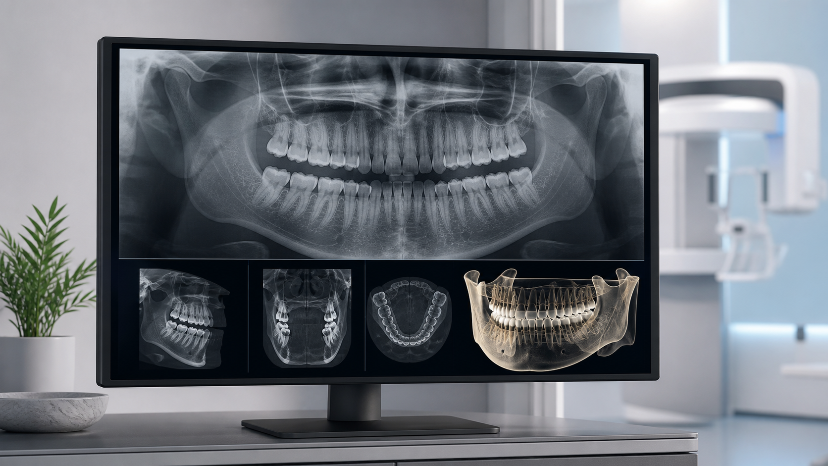
Key points in one minute
Key point: imaging is useful when it answers a specific clinical question and can change a dentist’s decision
- A small intraoral X-ray examines a limited area, a panoramic image gives an overall view of both jaws, and CBCT provides three-dimensional data for a selected region.
- An image complements rather than replaces a conversation and examination; it is chosen for expected diagnostic value, not simply by routine.
- X-ray examinations require justification and optimisation: the smallest suitable field and exposure are used to obtain diagnostic-quality information.
- Tell the team about pregnancy or possible pregnancy, childhood age, recent imaging and relevant health information before the examination.
What dental X-ray imaging is
X-rays pass through tissues differently according to their density, and a detector records an image. This can help a dentist assess roots, canals, supporting bone, contacts between teeth, tooth position and some changes that are hidden from view. A digital image is not a diagnosis by itself: it is interpreted alongside an examination and your health history.
Imaging can clarify a complaint, support treatment planning or monitor treatment when it can make a real difference to care. It also has limits. It does not reveal every cause of pain, the condition of oral soft tissue or the quality of daily cleaning, so other assessment methods remain important.
- An image should answer a defined question, not act as a universal check-up.
- The same image may be useful for one patient and unnecessary for another.
- Keep earlier images and tell the dentist where and when they were taken.
Periapical X-rays, panoramic imaging and CBCT
A small intraoral or periapical X-ray covers a limited area, often a few teeth, their roots and nearby bone. It may be used to examine a local problem, trauma, roots or a stage of root-canal treatment. A panoramic X-ray, often called an OPG, provides a broad overview of both jaws and the dental arches, but is not a detailed examination of every individual area.
Cone-beam computed tomography (CBCT) produces three-dimensional data that can be reviewed in slices. It may be appropriate for complex implant planning, an impacted tooth, selected trauma or a difficult anatomical question. CBCT is not a ‘best scan just in case’: its field of view and settings should be limited to the expected diagnostic benefit.
| Method | Typical contribution | Important limit |
|---|---|---|
| Intraoral X-ray | A small area, roots and nearby bone | Does not survey the whole jaw |
| Panoramic image / OPG | An overall view of jaws and dental arches | Fine detail may be limited |
| CBCT | Three-dimensional slices of a selected area | Used when indicated; the field of view should be no larger than needed |
When imaging may help — and when it may not
The dentist decides on imaging after taking a history and examining you. It may help investigate hidden decay, inflammation near a root, a fracture, bone changes around teeth, delayed eruption, or selected surgical, orthodontic and restorative plans. The timing of follow-up imaging also depends on the individual risk and clinical objective.
Neither being pain-free nor having pain alone determines which image is appropriate. FDA and ADA guidance emphasises that radiographs are selected when the additional information is expected to affect care. Show the dentist any suitable earlier images; they can sometimes prevent unnecessary repeat imaging.
Safety, exposure and image quality
X-ray imaging uses ionising radiation, so benefit and potential risk are considered for the individual patient. International and professional guidance uses justification and optimisation: an examination is performed for a medical question and with the lowest exposure that still provides adequate diagnostic quality. Digital equipment does not remove those principles.
Movement, positioning, metal restorations, anatomy and the selected field of view can affect image quality. Artefacts may hide a detail or resemble a finding, so a dentist may need to compare the image with the examination or occasionally repeat a limited image. Tell the team about pregnancy or possible pregnancy, a child’s age and recent imaging so the procedure can be planned safely.
- Ask which question the image is intended to answer and whether it may affect treatment.
- Ask whether existing images can be used.
- Do not request CBCT merely because it is 3D.
- For children, individual selection and the smallest adequate field are especially important.
Preparing for a consultation
Routine dental imaging usually needs no special home preparation. Bring or send earlier images and reports if available. Explain what is troubling you, when it began, whether there was trauma, what treatment has already been done and whether pregnancy is possible. These details help the dentist choose an appropriate examination without avoidable repetition.
After imaging, ask what is visible, what conclusions are supported and what remains uncertain. If treatment depends on an image, ask about alternatives, limits and the next step. Severe pain, a quickly increasing swelling, facial trauma, fever, difficulty swallowing or breathing require urgent in-person medical assessment rather than waiting for an article or an image to provide an answer.
- What clinical question will this image answer?
- Is a smaller field or an existing image sufficient?
- Which findings need treatment, monitoring or further clarification?
- When and why might follow-up imaging be needed?
Save before your visit
Questions worth asking your dentist
- Which type of image addresses my symptom or treatment stage?
- Can earlier imaging be used for comparison?
- Why has an intraoral X-ray, OPG or CBCT been selected for me?
- Which limits of this image matter for my treatment plan?
Sources
- Dental radiology - X-rays — International Atomic Energy Agency
- X-Rays/Radiographs — American Dental Association
- Medical X-ray Imaging — U.S. Food and Drug Administration


