
 23 Tumanyan St, Yerevan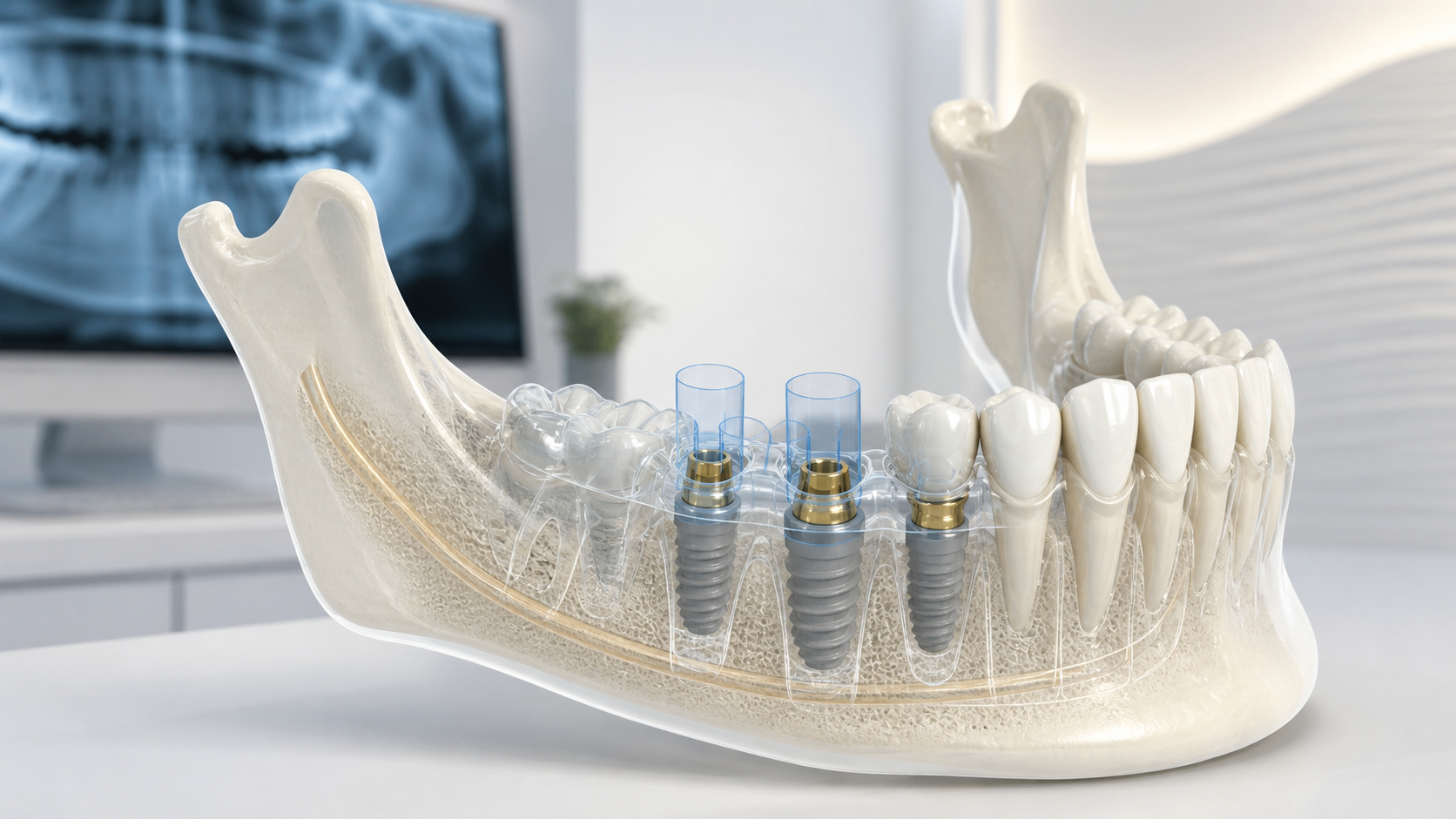
Key points in one minute
The plan makes treatment negotiable and safer, but does not turn it into a remote service
- Planning begins with an in-person collection of complaints and medical history, examination of teeth, gums and bite; one digital image does not establish a diagnosis.
- CBCT can show three-dimensional anatomy of the site, but is ordered based on clinical need and has limitations in accuracy and radiation exposure.
- The position of the implant is planned taking into account the future crown, adjacent teeth, soft tissues, bone and important anatomical structures, and not just free space.
- Template and digital plan - tools for transferring the solution; they do not override screening during treatment and do not guarantee the same outcome for everyone.
Short answer: what exactly are they planning?
Implantation usually consists of several decisions rather than a single action. The doctor evaluates whether it is possible to save your own tooth, where the future crown should be located, whether there is enough tissue to support it, and how the intervention will affect the neighboring teeth and bite. If implantation is suitable, the plan helps determine the sequence: first treatment of inflammation or caries, tissue preparation if necessary, then implant placement and subsequent prosthetics.
Planning makes the discussion more specific. Instead of a general proposal to place an implant, you can ask which problem the option addresses, what evidence supports it, what may change the timing and which alternatives exist. The decision follows an in-person assessment and informed consent; this article cannot replace those steps.
What data is collected before a decision is made?
First, they discuss the reason for contacting, diseases, medications taken, smoking, previous operations and expectations from the result. Then the teeth, gums, hygiene, mobility, space for a future crown and characteristics of jaw closure are assessed. This information may indicate that the first priority will be to treat active inflammation, save the tooth, or stabilize the gums, rather than getting an immediate implant.
Imaging is selected according to clinical need. A panoramic or intraoral radiograph may form part of the assessment, while CBCT provides a three-dimensional view of the area, including bone height and width, important canals, sinuses and adjacent roots. International guidance emphasises an individual justification for CBCT and the smallest suitable field of view. Imaging is always interpreted alongside clinical findings.
| Stage | What problem does it solve? | What it doesn't promise |
|---|---|---|
| Interview and inspection | Identify complaints, risks and tissue condition | Accurate forecast without additional data |
| Photos and scanning | Fix the shape of teeth, smile and bite | Assessment of hidden bone and neural structures |
| X-ray or CBCT as indicated | Assess site anatomy and plan safety | Zero error or no risks |
| Digital plan and template | Agree on treatment plan and transfer | Automatically successful operation or the same result |
Why start with the future tooth
The implant is a support for the future structure, so the plan is often built “from the crown back.” The doctor considers where the chewing tubercle should be, how the area will be cleaned, how much space there is between the teeth, and how the load will be distributed. Then checks whether the bone and anatomy allow the support to be placed in a safe position. Sometimes the aesthetically desired position does not coincide with the available volume of tissue - then a change in plan, tissue preparation, or another method of restoration is discussed.
Digital impressions and photographic protocols can help connect dental and soft tissue information with CBCT data. This is useful for explaining options, making a temporary structure or surgical guide. But digital imaging doesn't see everything: data quality, patient movement, artifacts, and software differences affect the model. Therefore, clinical supervision remains part of the process.
- Which tooth or area is being restored and is it possible to save your own tooth?
- How the future crown will participate in occlusion and hygiene.
- Are there enough bones and soft tissues in the right direction?
- Where the nerve canal, sinus, roots of adjacent teeth and other important structures pass.
- Are preparatory steps, a temporary solution or an alternative type of prosthetics necessary?
Digital Plan and Surgical Template: Useful Boundaries
After combining the data, the doctor can virtually select the approximate dimensions and position of the implant. This base is sometimes used to make a surgical guide, a device that helps guide the intended direction during the procedure. Its applicability depends on the clinical situation, template support, quality of input data, and team experience; it is not necessary in every case.
ITI publications indicate that linear CBCT measurements are generally accurate enough for planning purposes, but in some situations the error may exceed one millimeter. Therefore, the plan takes into account the margin to significant anatomical structures, and during treatment the doctor checks the compliance of the real situation with the model data. Statements such as “navigation eliminates risk” or “template guarantees perfect installation” would be misleading.
Limitations, risks and alternatives
The plan may change if tissue conditions after treating inflammation, extracting a tooth or preparing the site differ from the initial assessment. Timing and predictability are affected by hygiene, gum disease, smoking, general health, bite and the ability to attend follow-up visits. These factors should support an individual risk discussion, including what can be improved before treatment and how follow-up will be organised.
Implantation is not the only way. Depending on the cause of loss and tissue condition, tooth conservation with treatment and a crown, a bridge, a removable denture, or a deferred solution with a temporary option may be discussed. Each method has its own requirements for adjacent teeth, tissues, care, timing and risks. Making an informed choice is about comparing options, not about competing between implant brands.
How to prepare for a consultation and when not to wait
Take existing photographs and extracts, but do not do a new study just because you were asked to send it online. Talk about chronic illnesses, allergies, pregnancy, medications, and previous dental experience. It is useful to write down in advance what is more important: saving the tooth, restoring chewing, timing, appearance, ease of care or budget. The final conditions and scope of treatment are discussed only after assessing the specific situation.
Rapidly increasing swelling of the face or neck, fever, pus, jaw injury, uncontrolled bleeding, or difficulty opening the mouth, swallowing or breathing requires urgent medical attention. Pain, an unpleasant odour, bleeding gums, tooth mobility or a broken crown also warrants an in-person examination rather than self-diagnosis from an article.
Save before your visit
Questions worth asking your dentist
- Is it possible to save my own tooth in my situation and how does this option differ from implantation?
- What studies are really needed, what problem does each solve, and are there sufficient images already available?
- What will the whole path look like: preparation, installation, temporary and permanent construction, control?
- What factors in my health and care may change the plan, timing or risks?
- Which alternatives can be compared based on function, care, and limitations?
Sources
- Accuracy of Linear Measurements on CBCT Images Related to Presurgical Implant Treatment Planning — International Team for Implantology
- Computer-Guided Implant Surgery — International Team for Implantology
- Use of cone beam computed tomography in implant dentistry: consensus report — PubMed / Implant Dentistry
- Evidence-based clinical imaging diagnostic guideline for implant planning — PubMed / Imaging Science in Dentistry


