
 23 Tumanyan St, Yerevan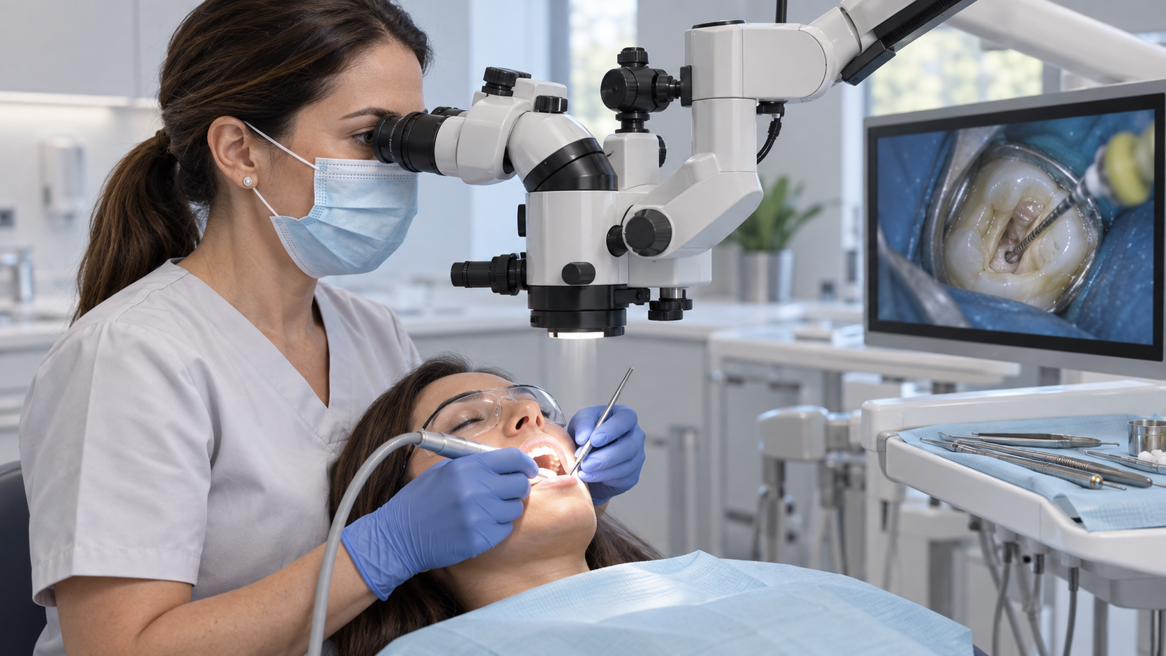
Key points in one minute
What the microscope can and cannot do
- Magnification improves the view of complex anatomy, but does not replace examination, imaging, canal cleaning or a final restoration.
- It can be useful when locating an extra canal, retreating a tooth or assessing a broken instrument or a perforation in the root wall.
- A microscope cannot promise success; conversely, its absence alone does not establish that treatment was inadequate.
- Contact your dentist about worsening pain, swelling or a lost temporary filling. Difficulty swallowing or breathing needs urgent medical care.
What does treatment under a microscope mean?
A dental operating microscope combines optical magnification with light directed along the clinician's line of sight. This can reveal small landmarks inside the tooth and in the accessible part of a canal. It does not make the entire root transparent: bends and deeper areas still require imaging, working-length measurements and clinical judgement.
A root canal is the space inside a tooth's root that contained the pulp. When the pulp is irreversibly inflamed or infected, the clinician removes affected tissue, cleans and shapes the canals, seals them and restores the tooth. The microscope makes details of this work easier to inspect; it is not a separate disinfection treatment.
Which problems can magnification help with?
An extra canal can be difficult to find, particularly when its opening is tiny or narrowed by calcification. If missed, part of the canal system may remain untreated. During retreatment, magnification can help the dentist examine old filling material and look for unusual anatomy. It may also help assess a crack, the edge of a root-wall perforation or the visible part of a separated instrument.
Seeing a problem does not mean it can always be corrected safely. Removing a fragment may require taking away root structure and could weaken the tooth; bypassing it or monitoring may be more appropriate in some cases. Whether a perforation can be repaired depends on its site, size, timing and the surrounding tissues. A tooth must also be restorable after canal treatment.
| Situation | What may become clearer | What else matters |
|---|---|---|
| Hidden or narrow canal | The opening and access landmarks | Imaging, root anatomy and safe access |
| Retreatment | Old material and a potentially missed canal | Cause of the previous problem and restorability |
| Separated instrument | The visible fragment and possible access | Risk of weakening the root during removal |
| Perforation or crack | Edges of a defect within view | Location, extent and the prospects for restoration |
What happens at the consultation and during care?
Your dentist will ask about symptoms and earlier treatment, then examine the tooth, gums and existing filling or crown. Focused dental radiographs are usually part of the assessment. Three-dimensional imaging may be considered when indicated and when ordinary views do not answer the question. A crack, periodontal condition or insufficient tooth structure can change the plan even if the canals can be reached.
If root canal treatment is appropriate, the clinician establishes access and isolates the working area, cleans and shapes the canals, then seals them. The tooth needs a well-sealed permanent restoration afterwards; some cases involve more than one appointment. The technique and timing depend on the individual tooth. Follow-up considers symptoms, function and imaging at intervals agreed with your clinician.
- What diagnosis do the examination and images support?
- What specific problem would magnification address in my case?
- Can the tooth be reliably restored after canal treatment?
- How would the plan change if a crack or inaccessible canal is found?
Limits, alternatives and what counts as success
Even meticulous canal work cannot rule out future infection. Magnification cannot compensate for a leaking restoration or a vertical root fracture. Comparative evidence isolating the microscope's effect on the long-term outcome of routine non-surgical root canal treatment is limited. Studies of endodontic surgery should not be applied automatically to every root canal case. A promise to save a tooth solely because a microscope is used is therefore unsound.
Depending on the diagnosis, options may include initial treatment, non-surgical retreatment, endodontic surgery, monitoring in selected circumstances or extraction with a discussion of replacement. A deep fracture or a tooth that cannot be restored may rule out retention. Success is judged by relief of symptoms, useful function, healing seen on follow-up images and the condition of the final restoration, rather than by the name of the device used.
Recovery and signs that need attention
Some tenderness, including discomfort on biting, can occur during the first few days. Your clinician should give instructions tailored to your case and explain when the tooth needs its permanent restoration. Protect a tooth with a temporary filling and follow the advice you were given. Do not choose antibiotics or pain medicines based on an article, and do not postpone final restoration without discussing it with your dentist.
Contact your treating dentist if pain worsens or persists, swelling develops, previous symptoms return, your bite feels different or the temporary filling is lost. Fever with increasing swelling, trouble opening your mouth, swallowing or breathing calls for urgent in-person medical assessment. If you are visiting Yerevan for treatment, agree a follow-up plan and obtain copies of your images and records before you travel onward.
- Which sensations are expected after my particular procedure?
- When should the permanent restoration and follow-up imaging take place?
- Which symptoms mean I should contact you before the scheduled visit?
Save before your visit
Questions worth asking your dentist
- Does every root canal procedure need a microscope?
- Can a microscope show the entire root?
- Does magnification guarantee that the tooth will be saved?
- What should I do if swelling appears after treatment?
Sources
- Microscopes in Endodontics — American Association of Endodontists
- Use of Microscopes in Endodontics: General — American Association of Endodontists
- Endodontic therapy using magnification devices: a systematic review — Journal of Dentistry via PubMed
- Endodontic Retreatment Explained — American Association of Endodontists
- Root Canal Post Treatment Care — American Association of Endodontists
- Ստոմատոլոգի մասնագիտական բնութագիրը — ՀՀ առողջապահության նախարարություն


