
 23 Tumanyan St, Yerevan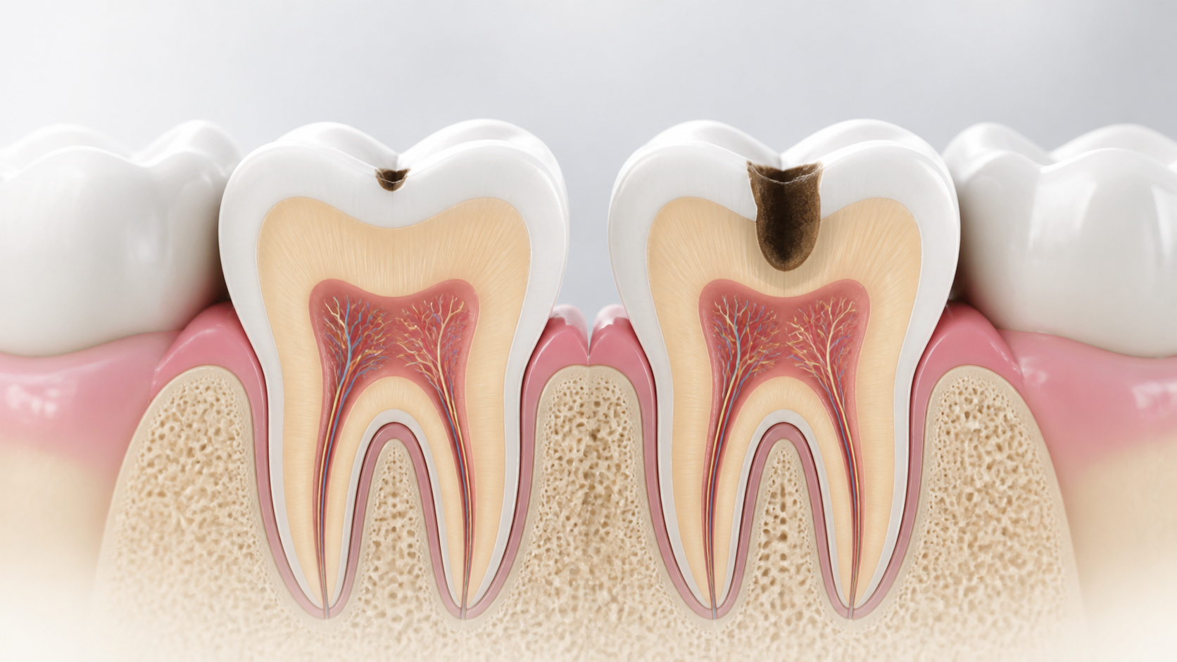
Key points in one minute
The main thing: early caries does not always hurt, and the amount of help depends on the depth and activity of the lesion
- Caries develops when acids formed in plaque from free sugars upset the balance of demineralization and restoration of tooth tissue.
- White or brown spot, tenderness, food entrapment, and cavity are possible but not required signs; cavities between teeth may not be noticeable without examination.
- Initial changes can sometimes be stabilized without drilling, and a cavity in the tooth tissue usually requires restorative treatment; the decision is made after diagnosis.
- Increasing pain, swelling, fever, difficulty swallowing or breathing is not a reason to wait for a scheduled appointment: an urgent medical assessment is required according to local regulations.
What is caries and how does it develop?
A biofilm, called plaque, constantly forms on the surface of the teeth. When free sugars from food and drinks frequently enter the oral cavity, microorganisms in the plaque produce acids. These acids temporarily reduce enamel mineralization. Saliva, fluoride-containing hygiene products and regular plaque removal support reverse recovery, but with frequent acid attacks, the balance shifts: the enamel area becomes more porous, then the process can move into dentin.
Therefore, tooth decay cannot be reduced to one “bad” product or to a lack of brushing on a single day. The risk is simultaneously influenced by the frequency of sugary snacks and drinks, hygiene, fluoride access, composition and quantity of saliva, fissure anatomy, existing restorations, habits and individual risk level. The World Health Organization lists free sugars and poor plaque removal as important modifiable factors.
Symptoms and stages: what you can notice
The initial mineral loss may appear as a dull white patch, especially after the tooth has dried out, and is often not painful. As the enamel changes, the stain may darken. If a cavity forms or the process extends to the dentin, there may be brief sensitivity to cold, sweet or sour foods, food entrapment, roughness, discoloration or chewing discomfort. However, the absence of symptoms does not exclude a lesion: for example, the contact surfaces between teeth are difficult to see independently.
The phrases “deep caries” and “nerve caries” are not a remote diagnosis. Pain can be associated not only with caries, and the degree of damage does not always coincide with how the tooth looks in the mirror. If the tissue near the pulp is inflamed or infected, the plan may include endodontic treatment; sometimes it is impossible to save a tooth. This is determined individually - after examination, tests and, if indicated, x-ray examination.
| What is noticeable | What could this mean? | Reasonable action |
|---|---|---|
| Matte white or dark spot without pain | Initial change in enamel or other cause of color change | Schedule an inspection, do not attempt to scrape or bleach the area yourself |
| Sensitivity to sweets or cold | Caries, dentin exposure, crack or other problem | Sign up for diagnostics; do not select treatment based on advice from the Internet |
| Cavity, chip or food keeps getting stuck | The integrity of the tissue or restoration is likely compromised | Contact your dentist as soon as possible |
| Increasing pain, swelling, temperature | Possible complication requiring prompt evaluation | Seek emergency dental or medical care |
How is a diagnosis made and why the same image is not always needed
Diagnosis begins with a conversation about complaints, habits, past restorations, nutrition, care products and general health. Then the doctor examines the cleaned and dried teeth in good light, assesses the surface, contacts between teeth, gums, old fillings and the risk of new lesions. It is important to distinguish an active carious lesion from an inactive stain, staining, enamel defect or restoration problem - tactics in these situations vary.
X-ray examination is used according to indications: it can help to see contact surfaces and assess the depth of the lesion, but does not replace a clinical examination and is not automatically prescribed for each symptom. The American Dental Association describes a systems approach: identifying the lesion, assessing its activity and individual risk, and then jointly deciding on preventive and treatment steps. Ask during your consultation what information the study will provide specifically for your situation and whether it will change the plan.
- Which signs indicate focal activity and which indicate an old stable change?
- Is the image needed now, what zone does it evaluate and how will the result affect the decision?
- What factors increase my risk of new lesions: frequent snacking, dry mouth, hygiene, restorations, or other?
- Which option will preserve the maximum amount of healthy tissue and how will control be organized?
Treatment: from early lesion control to restoration
If the surface has not yet been destroyed, the doctor may suggest observation, correction of risk factors and non-surgical methods aimed at stopping or slowing down the process. This is not “do nothing”: you usually need a clear plan for home hygiene, changes in sugar frequency, professional recommendations for fluoride products and a follow-up appointment. The approach depends particularly on age, caries risk, location and activity of the lesion. Not all white spots are reversible, and not every lesion is safe to observe without treatment.
When a cavity has formed or the tissues have softened so much that they cannot be preserved, the affected tissue is removed to the extent determined by the doctor, and the shape and contact are restored with a filling or other restoration. The modern approach tries to preserve healthy tissue, but does not promise the same method for all teeth. Deeper lesions may require pulp protection or root canal treatment; in case of a crack, severe failure or poor prognosis, alternatives are discussed. The material, number of visits, anesthesia and prognosis depend on the actual clinical picture, and not on the name of the procedure in the price list.
After treatment, the risk does not disappear: a new lesion may appear at the edge of the restoration and on other teeth. Therefore, the result is maintained with regular care and follow-up visits. If you live in Yerevan, are visiting from another region, or are planning treatment during a short trip, discuss in advance where and when the control will take place - especially if the plan involves several stages.
What you can do at home and when you need urgent help
Home prevention is not built around strict prohibitions, but around regularity. Brush your teeth twice a day with a fluoride toothpaste, clean between your teeth with a suitable toothpaste, and reduce the frequency of foods and drinks with added sugars between meals. Drinking water instead of sugary drinks and routine examinations help to notice changes in time. Children, pregnant women, people with dry mouth, braces, a large number of restorations or frequent new lesions need a personalized plan; fluoride concentration and additional medications are discussed with your doctor.
Make an appointment without delay if the pain recurs, gets worse at night, has a cavity, chipped tooth or restoration, swollen gums, changes in bite, bad taste, or difficulty eating. Rapidly increasing swelling of the face or neck, fever with worsening general condition, difficulty opening the mouth, swallowing or breathing are signs that require urgent medical attention according to local regulations. This article is intended to inform, but does not replace, examination and does not allow diagnosis from photographs.
Save before your visit
Questions worth asking your dentist
- Is the lesion active and what part of the tooth does it affect?
- What options are possible without drilling, and when is restoration really necessary?
- Is a photograph necessary and what information will it provide in my case?
- What habits and care products will actually reduce my personal risk of new lesions?
Sources
- Oral health — World Health Organization
- Sugars and dental caries: WHO technical note — World Health Organization
- Caries Risk Assessment and Management — American Dental Association
- Prevention and treatment of dental caries with mercury-free products and minimal intervention — World Health Organization


