
 23 Tumanyan St, Yerevan
Key points in one minute
What patients should know
- AI supports image interpretation; diagnosis and treatment decisions also require clinical assessment.
- Errors work both ways: software can miss an abnormality or flag a healthy area.
- Authorization of a particular device in another country does not establish its availability or permitted use in Armenia.
- Robotic guidance, tissue regeneration and digital twins serve different purposes and have different levels of readiness.
What dental AI actually does
An image-analysis algorithm looks for patterns learned from examples. Some systems flag areas that resemble decay or changes around a root; others identify fillings and crowns, assist measurements or help produce a report. These are distinct functions. A system that performs one task is not automatically validated for every diagnostic question.
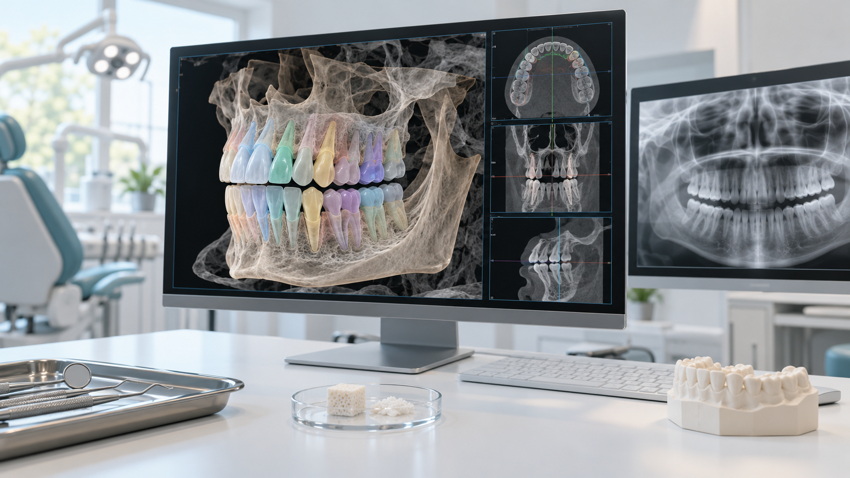
Segmentation means outlining teeth, bone and other structures within an image. It can make a three-dimensional model easier to use for planning, but an incorrect boundary can affect subsequent measurements. Automatically generated models need checking: an attractive visualization is not evidence that every detail is correct.
Dedicated diagnostic imaging software differs from a general conversational chatbot. A fluent chatbot response does not establish that the tool has been validated for that image type. Uploading medical images to a public service for self-diagnosis carries risks of both misleading advice and disclosure of sensitive information.
From a software prompt to a clinical decision
A safe process starts with symptoms, history and examination. The dentist requests appropriate imaging, interprets it and checks software prompts against the overall clinical picture. The exact workflow must follow the product instructions. Some tools are specifically intended as a second reader after the clinician has completed an initial assessment.
For example, a dark area on an image alone does not establish whether a process is active, explain pain or determine whether intervention is needed. Comparison with earlier images, examination of the tooth surface and gums, and additional clinical tests may be necessary. This is a general illustration of decision-making, not a patient case.
The dentist may accept a marker, dismiss it or investigate further. What matters to the patient is a clear explanation of why monitoring, prevention or treatment is recommended, what alternatives exist and what could change if care is delayed. Saying that the software made the decision does not replace that discussion.
Why AI misses problems or raises false alarms
A false positive is a flagged area that does not turn out to represent the target problem after assessment. A false negative is an abnormality the system misses. An unmarked image therefore does not guarantee health, and a colored box does not establish that a tooth needs drilling or extraction.
Errors can arise from overlapping anatomy, metal restorations, movement during imaging, insufficient sharpness or differences from training examples. Software designed for particular intraoral radiographs is not automatically suitable for a panoramic image or cone-beam computed tomography.
Data bias can occur when training and validation sets do not adequately represent the patients and equipment encountered later. Results from one device, age group or center cannot simply be assumed to apply everywhere. Software updates and changes in clinical workflow also matter.
People can make an additional error by trusting automation too much. A prominent marker may focus attention on one area, while a lack of prompts may create false reassurance. Maintaining independent clinical judgment reduces this risk without making the process infallible.
Making sense of accuracy claims
An accuracy figure means little without a defined task. Ask what findings were sought, which images were used, what reference assessment was applied and whether evaluation used data excluded from training. Sensitivity describes detection of existing abnormalities; specificity describes correct recognition of their absence. Neither is the probability of a diagnosis in an individual patient.
A separate question is whether the software helped clinicians in a clinical study and improved outcomes that matter to patients. Finding more abnormalities in a test image set does not automatically prove fewer complications, unnecessary procedures or costs. Long-term benefit requires studies designed to measure it.
ADA materials on AI standards emphasize image annotation, dataset quality and information protection. A standard sets out evaluation requirements; it does not itself establish the effectiveness of every product that mentions AI.
Regulatory status and questions in Armenia
Some systems have authorization for defined medical uses. For example, FDA document K210365 describes Second Opinion as a second-reader aid for certain intraoral radiographs of permanent teeth in patients aged 12 and older. This is an example from a 2022 document, not a universal description of every product version or all dental AI.
A US regulatory document concerns a particular device, version and intended use. It does not mean every feature from that brand has been validated for every patient, nor does it establish authorization or availability in Yerevan. Ask the clinic about the exact product, intended use, current documentation and applicable local requirements. This article does not claim that Full Clinic uses any of the named systems.
Armenian legislation on medical care addresses medical confidentiality and patients’ rights to information about their care. Before images are sent to an external service, ask who receives the data, for what purpose, where it is stored and how it is protected. Avoid posting images containing names or other identifiers in public chats. Removing a name does not always prevent identification.
Other technologies: clinical tools and research
Robotic guidance helps a surgeon plan and guide instruments. FDA documentation for the Neocis Guidance System describes assistance during dental implantation; it is not an autonomous dentist or evidence that every robot-assisted procedure is superior. Indications, team training, risks and alternatives need separate assessment. Overseas use does not establish availability in Armenia.
New biomaterials and regeneration cover many approaches. Existing restorative materials and specific tissue procedures should not be conflated with a promise to grow an entire new tooth. NIDCR describes research into scaffolds, cells, signaling molecules and tissue repair, with some approaches reaching clinical trials. The development stage must be established for each method.
A digital twin is a model linked to an individual’s data for analysis and simulation. An ordinary three-dimensional jaw image does not establish that software can reliably predict its future condition. Predictions require clinical validation, rather than convincing animation alone.
There is no single credible arrival date for a laboratory development in routine practice. Reproducibility, safety assessment, clinical trials, regulatory processes and professional training all take work. Some image-analysis tools are already used for defined indications; research technologies may evolve for years or never reach routine care.
| Technology | What to check |
|---|---|
| AI image analysis | Authorized task, image type, age group, independent validation and clinician assessment |
| Robotic guidance | Intended use, surgeon training, clinical evidence and alternatives |
| Biomaterials and regeneration | Specific material or method; laboratory versus clinical stage |
| Digital twin | What it predicts and how those predictions were validated |
A practical consultation checklist
If a dentist in Yerevan shows you an image with AI markings, ask for a plain-language explanation of each finding. Find out whether it is supported by examination and other evidence, what needs treatment now and what can be monitored. Discuss the pathway without AI as well: the absence of such software does not by itself indicate poor care.
For a complex or irreversible treatment plan, you can request copies of available medical records and discuss a second professional opinion. Visitors from the diaspora should also clarify the language of explanations, follow-up arrangements and how records can reach their next dentist. An impressive visualization alone is not a reason to consent to a procedure.
- What is the software called, and which task was it validated for?
- Is it suitable for my age and this image type?
- What has the dentist confirmed independently of its prompts?
- Will its output change treatment, and what alternatives are available?
- Will my data leave the clinic, and how is it protected?
When to seek help regardless of AI
Automatic image markers are not in themselves an emergency symptom. Urgency depends on the person’s condition. Tooth pain, swelling, pus or a suspected abscess warrant prompt dental attention; do not wait for an online interpretation of an image.
Difficulty breathing or swallowing, substantial swelling in the mouth, or eye or vision problems require emergency medical care. A lack of software warnings does not override these signs. This article provides general information; it does not diagnose, prescribe medicines or replace an examination.
Save before your visit
Questions worth asking your dentist
- Which finding needs attention, and is it supported by the examination?
- Can this change be monitored, and how will follow-up work?
- What evidence supports the proposed irreversible treatment?
- Can I obtain records for a second opinion?
Sources
- Second Opinion: FDA 510(k) K210365 — FDA
- Neocis Guidance System: FDA K161399 — FDA
- AI standards in dentistry — American Dental Association
- Ethics and governance of artificial intelligence for health — WHO
- Regenerative Medicine — NIDCR
- DOC Regenerative Medicine Program — NIDCR
- Armenian Law on Medical Care and Services for the Population — ARLIS
- Dental abscess — NHS


